The determination and interpretation of biological structures at the atomic level is an essential part of biopharmaceutical industry and life science R&D, with applications in:

Precise structural knowledge of a therapeutic target – drug interaction is crucial for any modern drug discovery and development approach.
  • It facilitates Hit-To-Lead (H2L) and Lead Optimization (LO) processes, enabling guided design using high-resolution protein-ligand structures, thus reducing the costs and timelines while increasing the chance of regulatory approval.
  • Additionally, structurally-enabled targets offer opportunities for novel hit identification (for example, by crystallographic fragment screening) or validation of hits identified by orthogonal methods.

Our holistic approach to structure determination allows us to choose the best method for any target, from single small proteins to complex intercellular arrangements.

High-resolution antibody-antigen structures provide detailed knowledge of the interaction between an antibody and its binding site on an antigen (epitope) at a single-residue resolution, which is necessary for the development of antibody-based biologics and diagnostic tools.
  • Understand the antibody-antigen interaction to enable further antibody development
  • Improve both the affinity and the specificity of your antibody without wasting resources on blind trial-and-error approaches
  • Validate the antibody-antigen complex integrity
  • Study proteins independently of labelling methods and cross-linking artefacts

Learn how our methods can boost your biologics development!

Bispecific antibodies (bsAbs) bind two epitopes at once, thereby bringing two different cells into proximity to elicit an immune response, as seen for example in cancer immunotherapeutics. Our services allow to study the bsAbs at various levels:
  • Confirm bsAb stability (often problematic) with Mass Photometry
  • Confirm individual Fab-antigen interactions by X-ray crystallography or Cryo-EM
  • Improve the affinity and specificity of the individual Fabs with structure-based engineering
  • Study the cell engagement of your bsAbs in near-native conditions with in situ Cryo-Electron Tomography (Cryo-ET)
The combination of these structural biology and biophysics methods allows for precise development of highly potent and selective antibodies with expected lower failure rates in clinical trials.
Protein engineering has been a cornerstone of Biotechnology research for decades. With the recent expansion of Biologics development, it has also become an important focus of the Biopharmaceutical industry. With the massive increase in knowledge of protein folding and function, it is now easier than ever to design and develop new proteins with tailored capabilities and resilience.

Structure determination is a crucial building block of this approach, enabling analysis of starting templates, rational design based on high-resolution structures, and verification of subsequent designs.

  • X-ray crystallography and Cryo-EM techniques provide high-resolution special information essential for informed design
  • Protein production and purification, followed by stability and homogeneity testing by Mass Photometry to ensure protein quality for downstream functional testing
Understanding how a pathogen interacts and affects a target host cell is key to the development of countermeasures such as vaccines or antibiotics. Structural biology enables insights into pathogen biology on various levels:
  • Host protein – viral protein interactions can be investigated with X-ray crystallography or Cryo-EM
  • Cryo-ET enables us to see pathogen processes directly in the cells and tissues, flash-frozen as they were happening

Learn more about the newest advances in host-pathogen research by Cryo-ET here.

Many modern therapeutics utilize delivery systems that bypass biological barriers to reach their desired place of action. Well known examples are RNA vaccines, cancer therapeutics, and gene therapy.

Structural biology approaches grant knowledge of the vector’s loading state, morphology, and distribution, which are all key to successful drug delivery vector development.

  • Negative staining EM is a cost-effective method to determine size distribution, morphology, and loading state of any large lipid- or capsid-based nanoparticle
  • Cryo-ET (electron tomography) images the drug carrier in situ, while it interacts with the target cells. Like no other experimental method, Cryo-ET enables understanding not only at the morphological level, but also on a functional level in the context of native environment.
AI-based protein structure prediction tools, such as AlphaFold, as well as protein design tools, have revolutionized the pharmaceutical industry by enabling rapid and accurate prediction and design of protein structures.

However, predictions remain only that until validated with experimental methods. Structural biology experiments ensure the accuracy and applicability of AI-predicted proteins in real-world scenarios, which is especially important in the area of Biopharmaceuticals.

  • Structure determination by X-ray crystallography or Cryo-EM allows verification of AI-predicted models by comparing computational and experimental data, identifying discrepancies, and refining predictions.
  • Integration of AI predictions with structural biology accelerates the identification of novel protein functions and interactions.
PROTACs (PROteolysis-TArgeting Chimeras) and Molecular Glues – emerging tools for targeted protein degradation – are currently redefining our attitude towards small molecule drug development.

By providing high-resolution insights into protein-protein interactions, ligand binding, and complex assembly, structural biology enables the rational design of these molecules.

X-ray crystallography and cryo-EM help visualize the ternary complexes formed between the target protein, the E3 ubiquitin ligase, and the PROTAC molecule. This allows researchers to optimize the linker length and orientation for efficient ubiquitination and subsequent degradation.

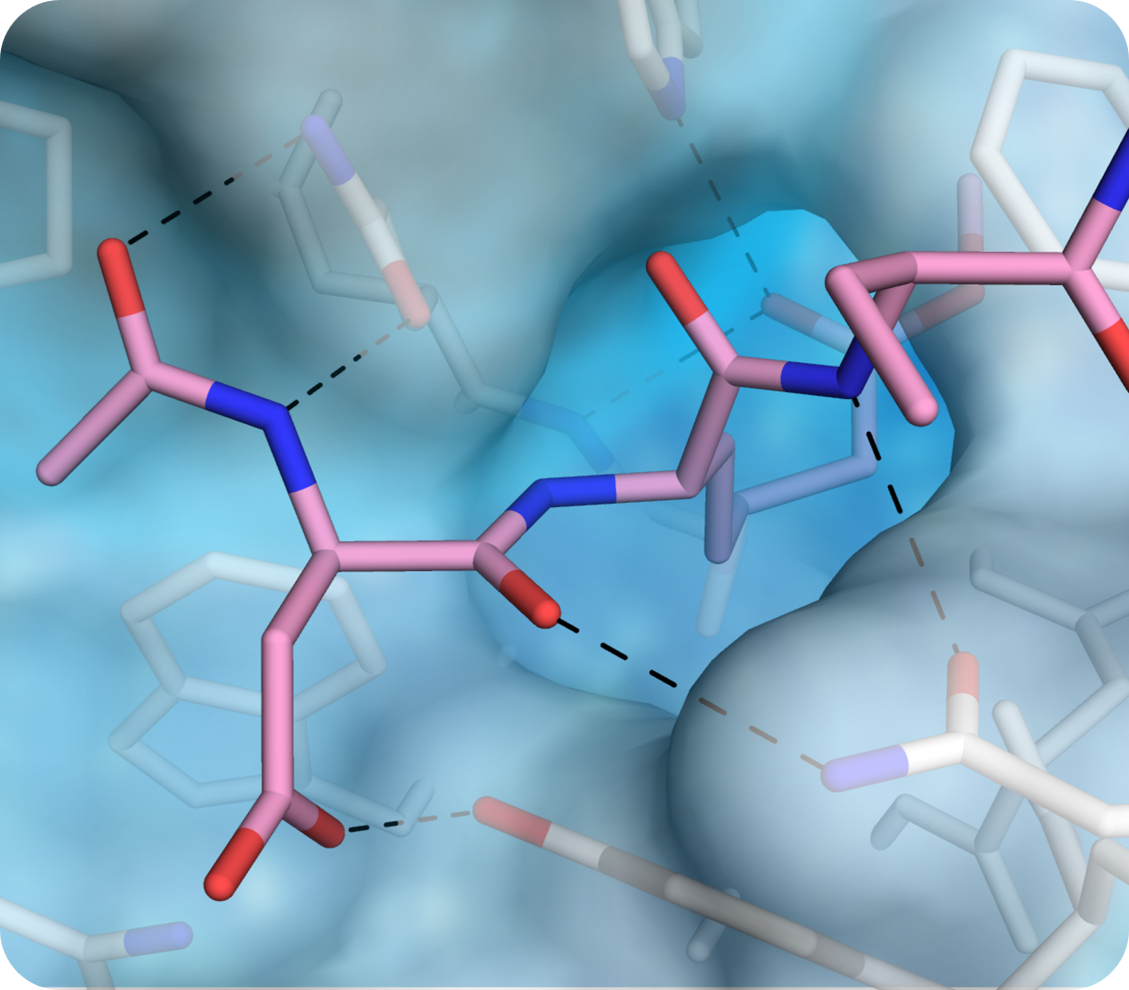
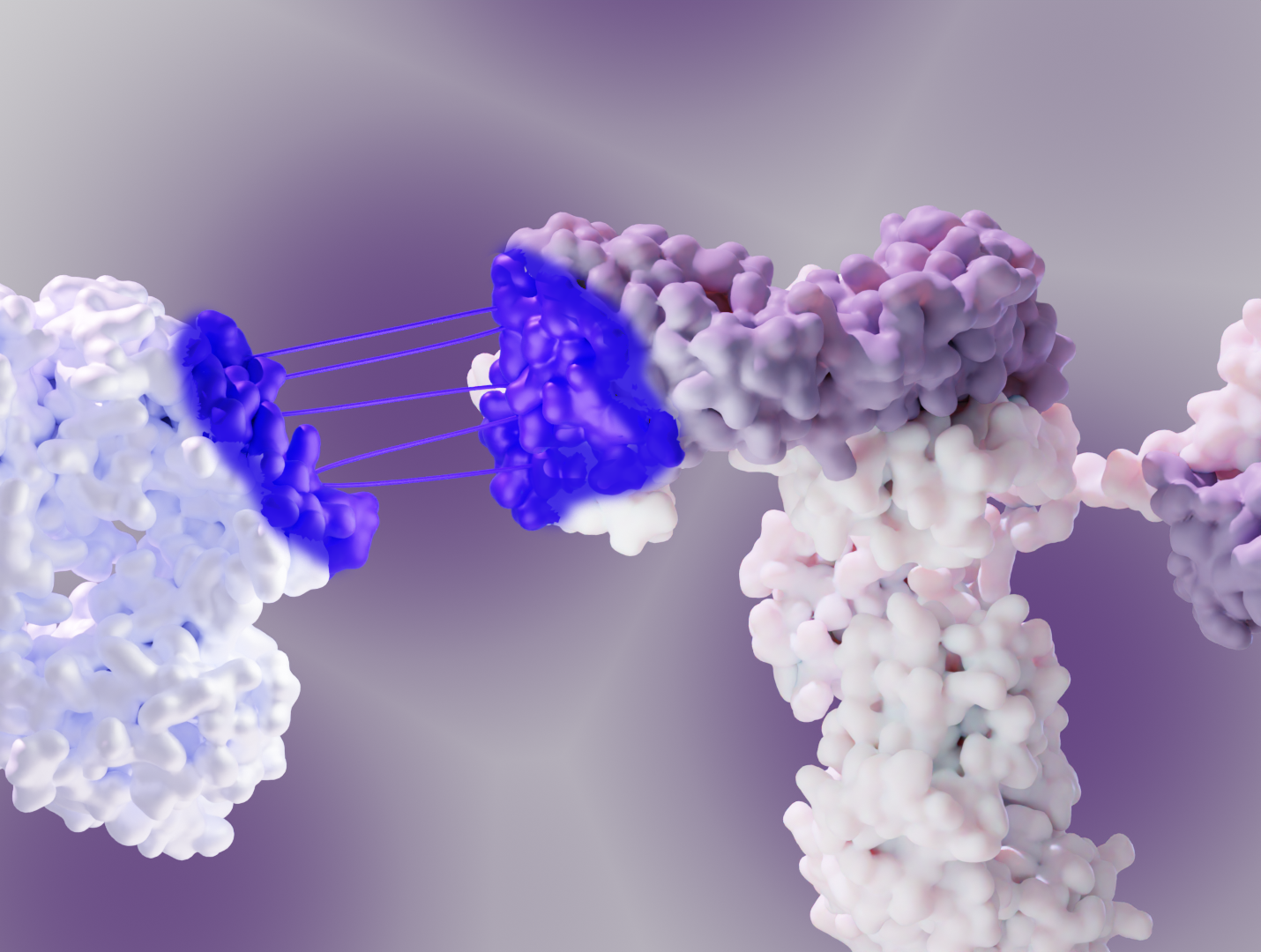
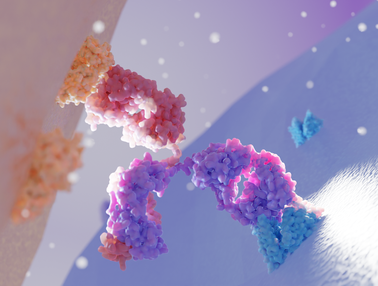
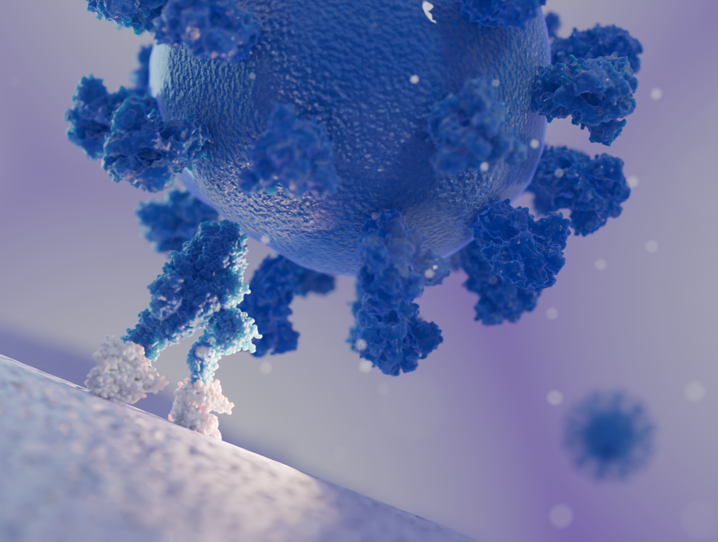
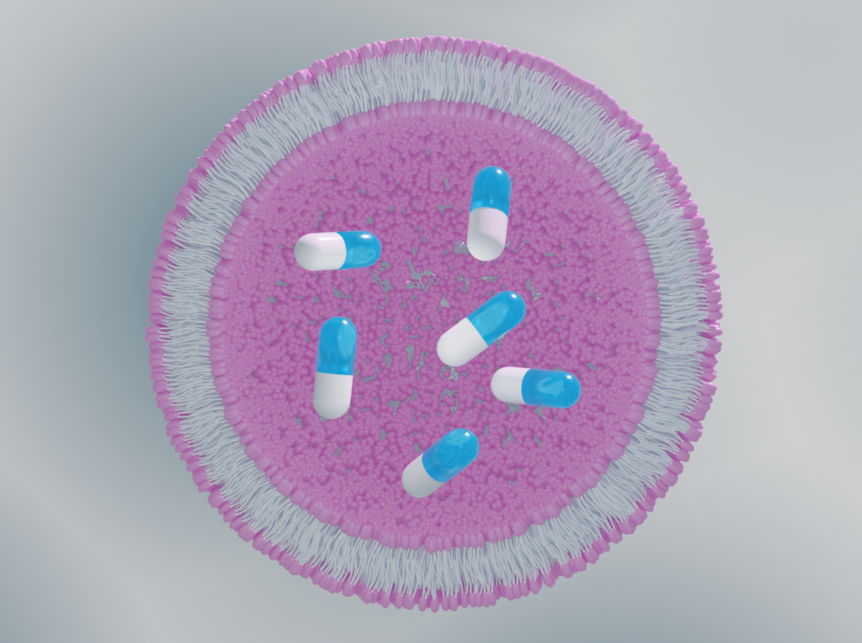
Stable, reproducible, and homogeneous samples are the cornerstone of every biochemical production from enzymes, through antibodies and antigens, to lipid nanoparticles and viral particles. 
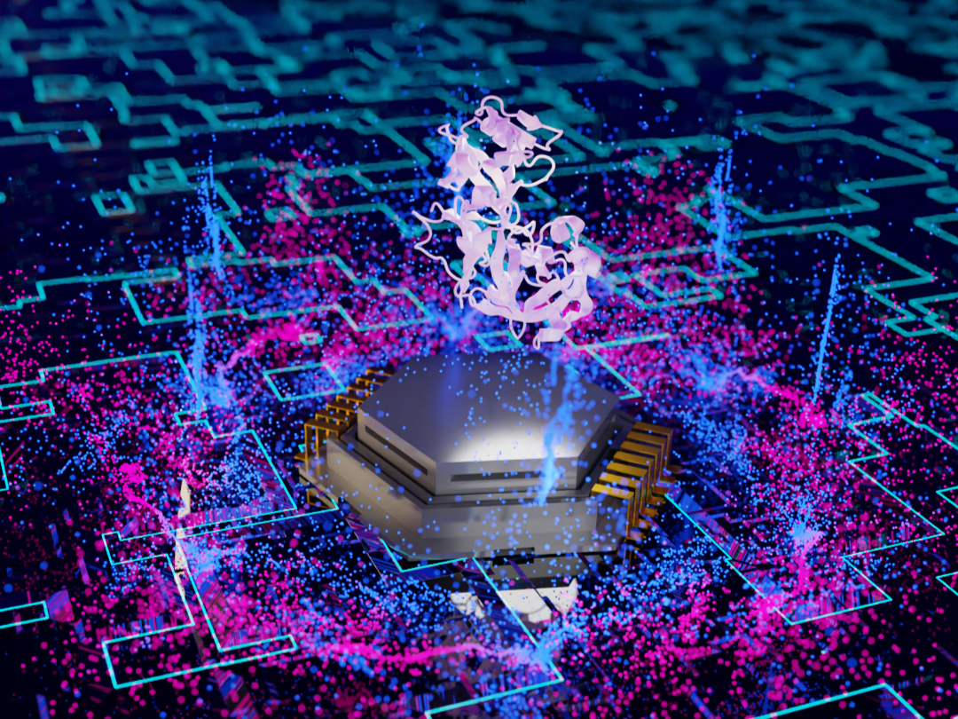
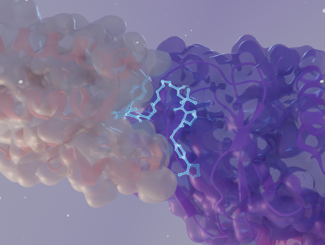
Our  versatile approaches utilize Structural Biology and Biophysics methods to ensure that highest quality samples are used in your studies and development pipelines.
  • Mass Photometry is a fast and cost-effective method that directly analyzes homogeneity, degrees of multimerization, and stability over time for samples such as membrane proteins, antibodies and ADCs, or AAVs
  • X-ray crystallography and single-particle Cryo-EM, unlike any other method prove the identity and quality of the sample via direct visualization
  • Negative staining EM provides quick and convenient way to analyze larger samples in terms of size and distribution, with particular use for AAV and nanoparticle characterization